
Case Report: Mandibular Full‑Arch Implant Restoration
I. Basic Information
Name: Ms. Zhai
Gender: Female
Age: 64 years
Chief Complaint: Difficulty in mastication in the mandible for several months; requesting oral examination.
Present Dental History: The patient reported having had weak biting force for six months, affecting food intake, and thus presented for consultation.
Past Medical History: Generally healthy; no significant findings; denies any systemic diseases.
II. Clinical Examination
Specialised Examination:
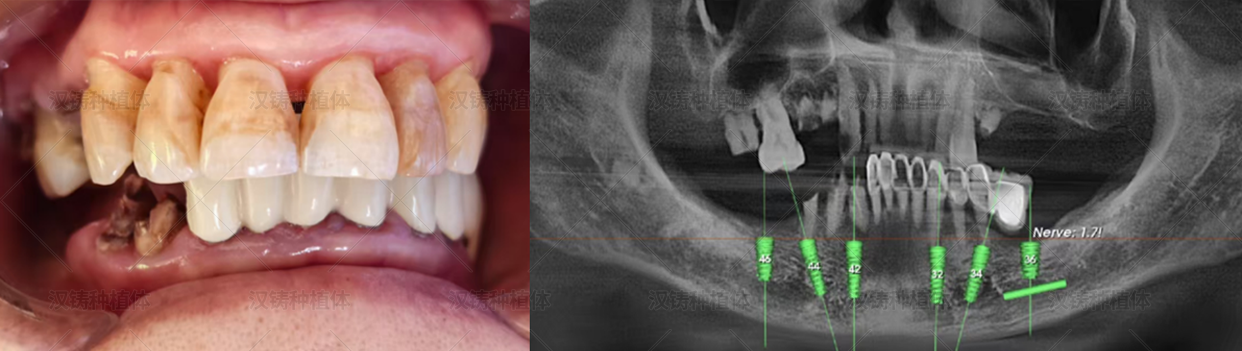
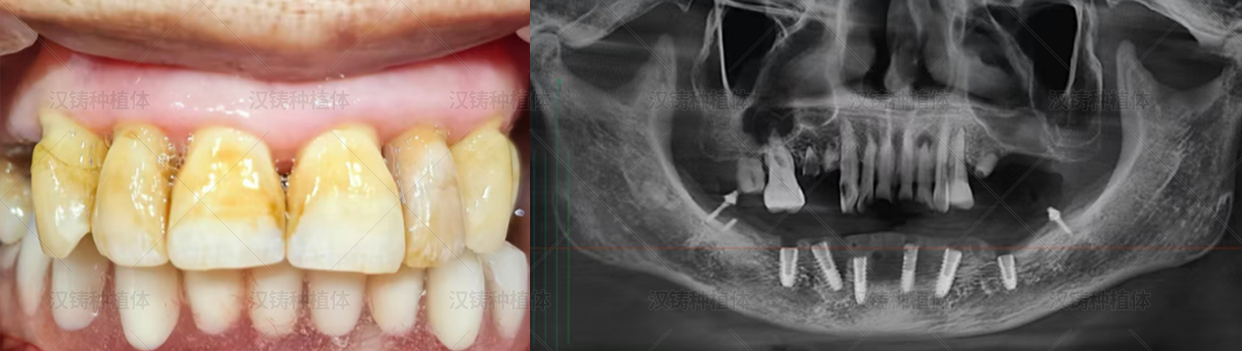
Defective restorations were noted on teeth #11, #12, #21, #22, #23, and #25, with gingival erythema and a dark red appearance, bleeding on probing, and deep periodontal pockets detected. Plaque index was 4; calculus index was Grade I (deposits not exceeding one‑third of the crown); abundant soft debris and mild halitosis were noted. These teeth exhibited Grade III mobility.
Teeth #43, #44, and #45 presented as residual roots with Grade III mobility, caries extending to the cervical region, and detectable root canal orifices. Gingival erythema, a dark red appearance, bleeding on probing, and deep periodontal pockets were noted. Plaque index was 4; calculus index was Grade I; abundant soft debris and mild halitosis were present. No other abnormalities were found.
Severe resorption of the mandibular alveolar bone was observed.
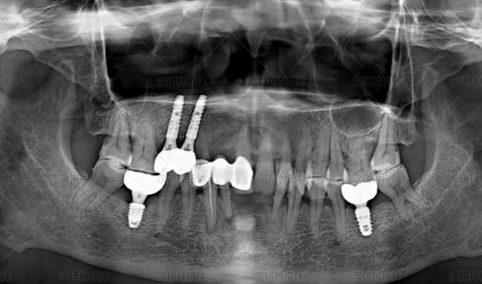
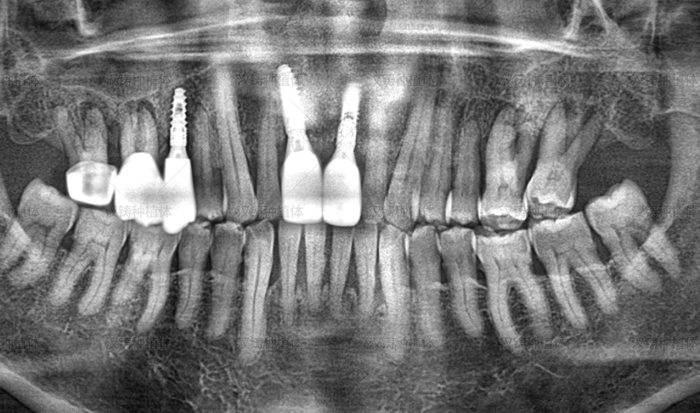
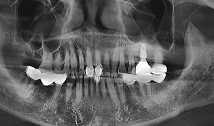
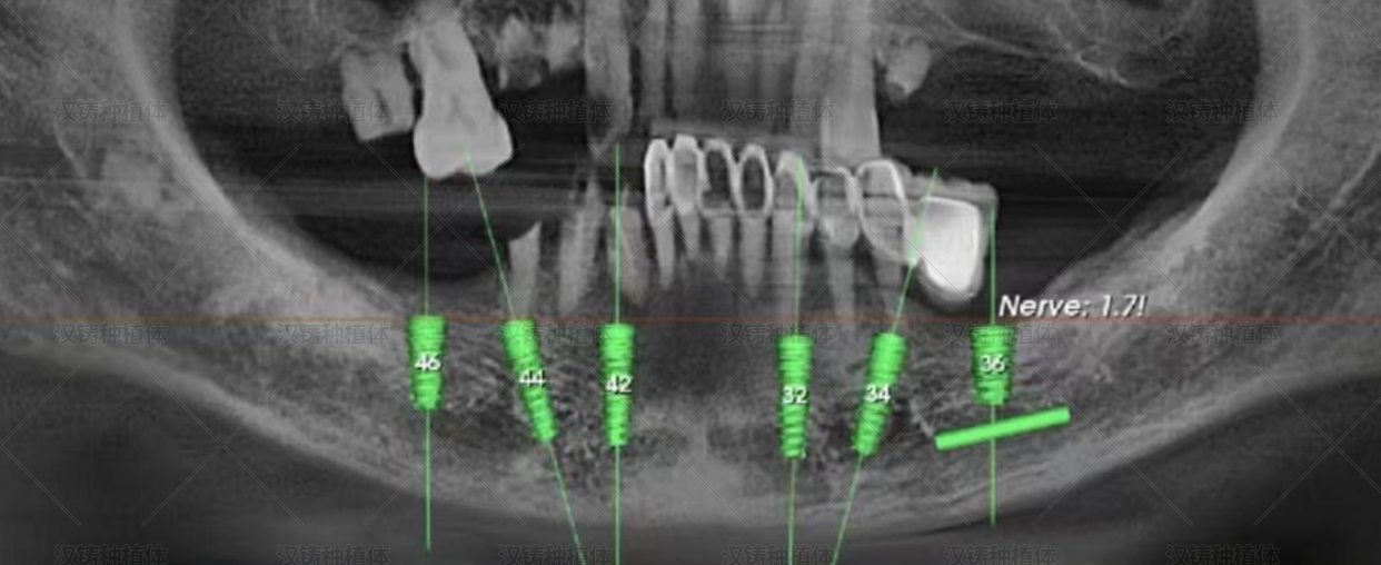
Auxiliary Examination: Radiographic examination was performed.

III. Diagnosis and Treatment Plan
Diagnosis:
#43, #44, #45: Residual roots with Grade III mobility.
#11, #12, #21, #22, #23, #25: Grade III mobility with defective restorations and moderate periodontitis.
Treatment Plan: Mandibular full‑arch extraction and implant placement (6 implants). Implant system: Hanzu®. Implant sizes and sites:
#33: 4.0×13 mm
#34: 4.5×13 mm
#36: 5.0×8 mm
#43: 4.0×13 mm
#44: 4.0×13 mm
#46: 5.0×10 mm
IV. Treatment Procedure
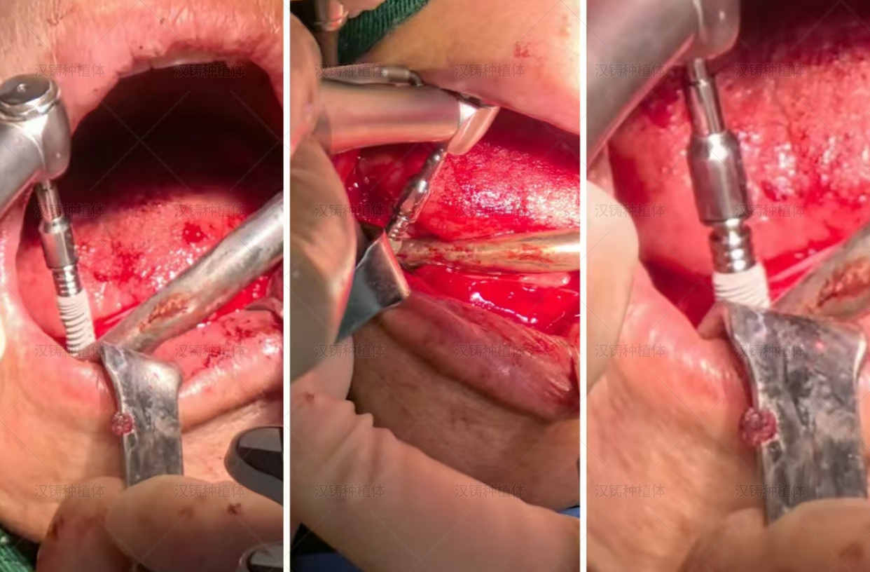
Surgical Phase (December 2024): The patient was fully informed of the risks and benefits and signed the informed consent form for implant restoration. Routine disinfection, draping, and local infiltration anaesthesia were carried out. After anaesthesia took effect, the teeth were extracted with forceps. A horizontal incision was made along the alveolar ridge crest, and a full‑thickness mucoperiosteal flap was reflected. The sites were localised, the extraction sockets were curetted, and any sharp bony edges were smoothed using rongeurs. The bone surface was prepared with a round bur, and osteotomies were prepared to the predetermined depths. Six Hanzu® implants were placed as planned:
#33: 4.0×13 mm
#34: 4.5×13 mm
#36: 5.0×8 mm
#43: 4.0×13 mm
#44: 4.0×13 mm
#46: 5.0×10 mm
Restorative abutments were connected, and telescopic crowns were seated and tapped into place. Radiographs confirmed satisfactory positioning. Tension‑free primary closure was achieved. Impressions were taken, plaster models were poured, and the casts were mounted on an articulator. A provisional removable prosthesis was fabricated extraorally, tried in, and adjusted to ensure patient comfort, completing the immediate loading protocol. The patient was prescribed antibiotics for 4–7 days and advised to avoid chewing hard foods. Post‑operative instructions included: no brushing or rinsing for 24 hours, a soft‑food diet at lukewarm temperature, maintain oral hygiene, and return for suture removal in 2 weeks. The patient was advised to return promptly if any discomfort occurred.
Prosthetic Phase (June 23, 2025):
Oral Examination: No significant erythema or swelling noted in the implant areas.
Auxiliary Examination: Radiographs showed good osseointegration with no abnormal findings.
Procedure: After confirming satisfactory bone‑implant integration on radiographs, the provisional prosthesis was removed with a crown remover. The oral cavity was cleansed, and the abutments were cleaned with 75% alcohol cotton pellets. Torque was reapplied to 35 N·cm. After confirming no abnormalities, an alginate impression was taken extraorally, bite registration was recorded, and plaster models were poured and mounted on an articulator. A definitive removable prosthesis was fabricated extraorally, tried in, and adjusted to ensure optimal fit and function. The final restoration was delivered.
Instructions: Maintain meticulous oral hygiene and return for regular follow‑up visits.
V. Case Summary
In the clinical experience of Director Wu Jiangbo, the Hanzu® implant system has exhibited strong initial self‑tapping ability and high primary stability, making it suitable for full‑arch implant restorations with immediate loading. It also performs well in a variety of bone conditions, including compromised bone quality, narrow or atrophic alveolar ridges, and other complex scenarios. The implant design takes into account individual variations, offering a wide range of sizes. It provides long‑term service life and high implant survival rates.
VI. Clinician Profile
Name: Wu Jiangbo
Affiliation/Title: Xiangrikui Dental Clinic, Jiyuan City, Henan Province
Honours and Achievements:
Director of Jiyuan Xiangrikui Dental Clinic
Visiting Scholar in Implantology, Department of Medicine, University of Mainz, Germany
Member of the Henan Provincial Oral Analgesia Committee
Member of the Chinese Stomatological Association
Areas of Expertise: Oral surgery, edentulous implantology, elderly prosthodontics, minimally invasive and pain‑free implant surgery.
Director Wu Jiangbo was born into a family of dentists. He has undertaken advanced training at the Fourth Military Medical University, the Implant Hospital of West China College of Stomatology, and the University of Mainz in Germany. He is a certified practitioner for the Bicon system (USA), the Isdian system (Korea), and the Nobel Biocare system (Sweden). With over a decade of dedicated practice and continuous education in advanced implant techniques, he has earned high recognition from patients with missing teeth.